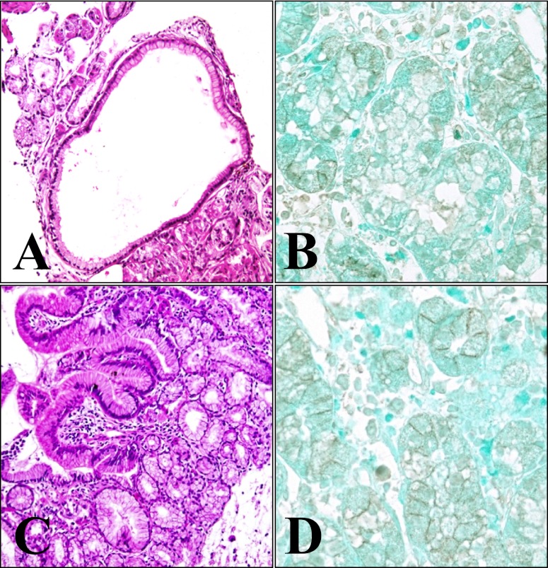
Fig. 2.
Histopathological features and immunohistochemical staining for β-catenin of case 1. Dilated glands lined by oxyntic epithelium are noted in the fundic gland polyp of the stomach (A, HE, ×100). The diffuse membranous and partly cytoplasmic distribution of β-catenin are noted (B, ×400). In the heterotopic gastric mucosa of the duodenum, the oxyntic epithelium is typically nondysplastic, and the junction between the gastric-type and duodenal surface epithelium is apparent (C, HE, ×100). β-catenin immunostaining shows mainly membranous staining (D, ×400).

