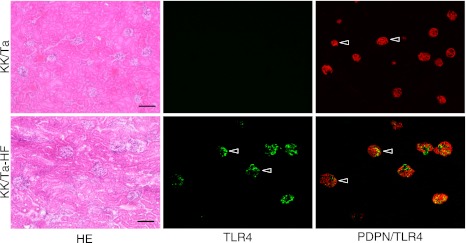
Fig. 4.

Immunostaining for TLR4 and podoplanin in the KK/Ta and KK/Ta-HF mouse kidneys. The immunostained sections were re-stained by the H-E staining. The HE staining shows that the kidney tissue is collapsed by edema and renal tubules have expanded in the cortex of the kidneys in KK/Ta mice which have been fed the high fat diet feed (KK/Ta-HF). In the KK/Ta mouse kidney sections, all glomeruli were immunostained with an antibody for the podocyte marker, podoplanin (PDPN), while there were no cells reacting with ani-TLR4. In the KK/Ta-HF mouse kidney sections, almost all podoplanin-positive glomeruli were immunostained by anti-TLR4 (arrowheads) whereas the Bowman capsules, distal and proximal tubules, collecting tubules, lymphatic vessels, and blood vessels outside glomeruli, were not immunostained. Bar=100 µm.
