
Fig. 7.
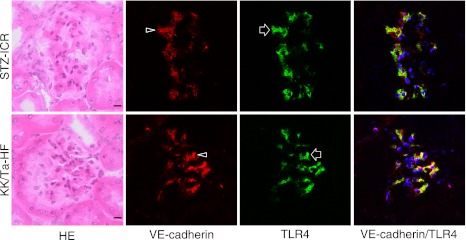
Laser-scanning confocal microscopy of immunostaining for VE-cadherin and TLR4 on the glomeruli of the ICR-STZ and KK/Ta mouse kidneys. The endothelial region reacting with anti-VE-cadherin (arrowheads) coincided with the region reacting with anti-TLR4 (arrows) in the merged images (rightmost panels). Bar=20 µm.