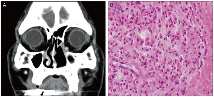
Fig. 2.
(A) Orbital computed tomography demonstrated an orbital mass located in the medial wall of the left orbit. (B) The recurrent tumor demonstrates the same morphology as the original tumor. The recurrent tumor is also composed of oncocytic epithelial cells with abundant eosinophilic cytoplasm (hematoxylin-eosin, ×400).

