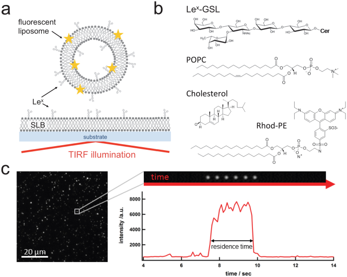
Figure 1. Scheme of the TIRF based approach.
(a) Schematic illustration of the detection principle. Fluorescently labeled liposomes containing Lex-GSLs interact with an SLB containing Lex-GSL. TIRF-based illumination is used to track surface-bound liposomes. (b) Chemical structure of lipids used. (c) A typical TIRF image of surface bound liposomes together with a kymograph and the intensity profile of a small image area containing a single liposome.

