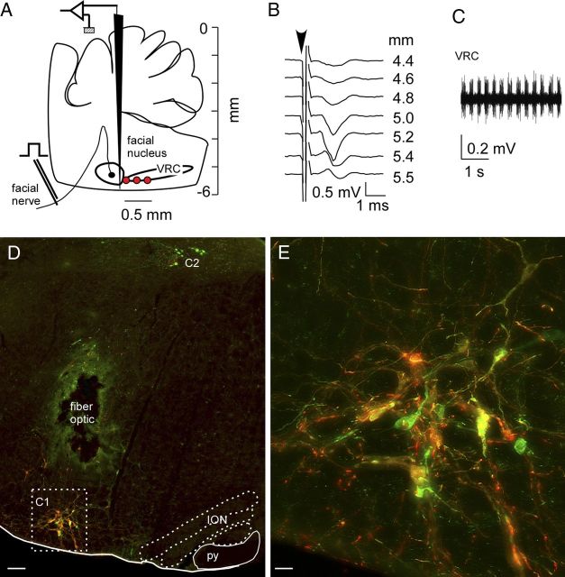
Figure 1.
Selective expression of ChR2-mCherry in RVLM-CA neurons in DβHCre/0 mice. A, The AAV2 vector was microinjected at the medial edge of the ventral respiratory column (VRC; red dots). These sites were identified electrophysiologically by their anatomical proximity to the facial motor nucleus. B, Example of antidromic field potentials recorded in the facial motor nucleus as the AAV2-containing injection pipette was lowered through the medulla oblongata. C, Multiunit respiratory activity recorded just caudal to the facial motor nucleus, further defining the AAV2 injection sites. D, Transverse hemisection through the left medulla oblongata of a DβHCre/0 mouse that received injections of AAV2-DIO-ef1α-ChR2-mCherry 4 weeks prior into the left RVLM. mCherry (red) and TH (green) are both detected by immunofluorescence. In the C1 region of the RVLM, a majority of the CA neurons expressed the mCherry transgene (yellow/orange). The dorsal group of CA neurons (C2) was not transduced. The lesion was caused by the insertion of the fiber optic, the green color surrounding the lesion is a histological artifact. ION, inferior olivary nucleus; py, pyramidal tract. E, enlargement of the C1 region outlined in D. Scale bars: in D, 100 μm; E, 20 μm.

