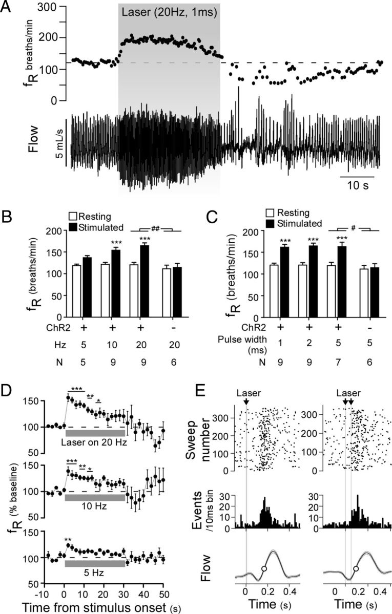
Figure 4.

Optogenetic activation of ChR2-expressing RVLM-CA neurons activates breathing in conscious DβHCre/0 mice. A, Plethysmography recording of the respiratory effects of photostimulation in an AAV2-DIO-EF1α-ChR2-mCherry-injected DβHCre/0 mouse (top, respiratory frequency (fR); bottom, respiratory flow signal, upward deflection in waveform represents inspiration). B, C, Relationship between initial change in fR and stimulation frequency at constant light pulse duration (2 ms) (B) and light pulse duration at constant stimulation frequency (20 Hz) (C) in experimental (ChR2-mCherry) and control mice (mCherry alone). ***p < 0.001 for resting versus stimulated in experimental mice by two-way ANOVA with multiple comparisons. #p < 0.05, ##p < 0.01 for the interaction of ChR2-mcherry expression and stimulation in experimental versus control mice. D, Time course of the change in fR during 30 s stimulation trials at 20, 10, and 5 Hz with a 5 ms pulse width (top to bottom traces, respectively) (N = 7). E, Two examples of photostimulus-triggered histograms plotting the onset of inspiration relative to light pulses delivered at low frequency designed to entrain respiratory rhythm. Both the raster plot and binned event counts (10 ms bins) indicate that single (left, 10 ms pulse at 2 Hz) and double (right, 5 ms pulses at 1.83 Hz) light pulses produced a clustering of inspiratory events shortly after the stimulus. Lower trace, Photostimulus-triggered average of respiratory flow during the entrainment protocol.
