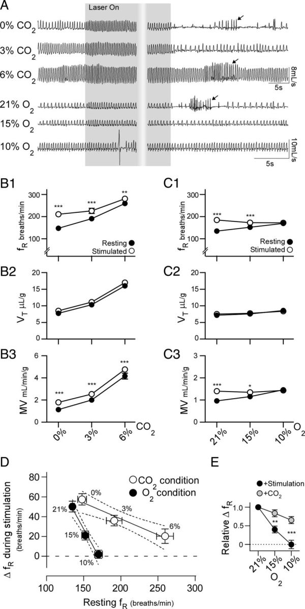
Figure 5.

Changes in breathing frequency during stimulation of RVLM-CA neurons is differentially occluded by hypoxia and hypercapnia. A, Initial respiratory effects of photostimulation (5 ms pulse at 20 Hz for 30 s) and poststimulus hypoventilation in a ChR2-mCherry-expressing DβHCre/0 mouse during hyperoxia (100% O2, trace 1), hyperoxic hypercapnia (3 and 6% CO2 balanced in O2, traces 2 and 3, respectively), and a different mouse during normoxia (21% O2 balanced in N2, trace 4) and poikilocapnic hypoxia (15 and 10% O2 balanced in N2, traces 5 and 6). Arrows identify bouts of poststimulus hyperventilation. Note that the period of hypoventilation following the offset of the stimulus under hyperoxic and normoxic conditions is largely abolished when respiratory drive is increased by activating the chemoreceptors. B, Group data of the effects of photostimulation on fR (B1), VT (B2), and MV (B3) under hyperoxic and hyperoxic hypercapnic conditions. N = 9 for each condition, *p < 0.05, **p < 0.01, ***p < 0.001 for resting versus stimulated condition. C, Group data of the effects of photostimulation on fR (C1), VT (C2), and MV (C3) under normoxic and hypoxic conditions. N = 7 for each condition *p < 0.05, **p < 0.01 for resting versus stimulated condition. D, X-Y plot of the average change in fR during stimulation in relation to prestimulus fR under each gas mixture presented in B1 and C1 (p = 0.0001 for difference between slopes, hypercapnia r2 = 0.44, hypoxia r2 = 0.73; error bars indicate 95% CI). E, Change in fR caused by RVLM-CA neuron stimulation and a brief CO2 challenge (15 s of 15% CO2) during graded hypoxia, normalized to the response fR under normoxic conditions. **p < 0.01, ***p < 0.001 for stimulation versus CO2 condition.
