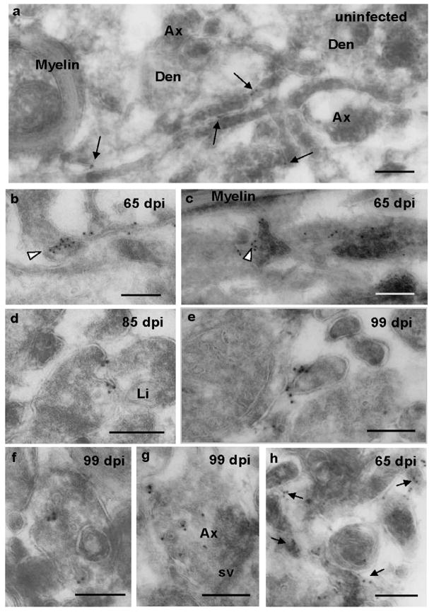
Figure 4. Cryo-immunogold EM of cell surface and vesicular PrP in stratum oriens.
Saf32 cryo-immunogold EM labeling of PrP on the cell surface (a–e, h) and vesicles (f, g) in stratum oriens (a-g), and alveus (h). (a) Sparse labeling of PrPC (arrows) in neuropil of uninfected hippocampus. (b–h) Clusters of labeling indicate PrPSc at 65 dpi (b, c, h), 85 dpi (d), and 99 dpi (e, f, g). PrPSc can be seen on very small diameter processes in (b, e), and at cell-cell junctions (b and c arrowheads; d, e). (c) Cryo-section, 200 nm thick, grazing the surface of a process that runs horizontally across the image. Several other processes, which appear darker, are on top of this. The arrowhead indicates Saf32 labeling at a point of contact. In (d), the plasma membrane of one of the processes appears to be invaginating. Arrows in (h) indicate clusters of labeling. Abbreviations: Den, dendrite; Ax, axon; sv, synaptic vesicles; Li, lipid droplet. Bars, 200 nm.

