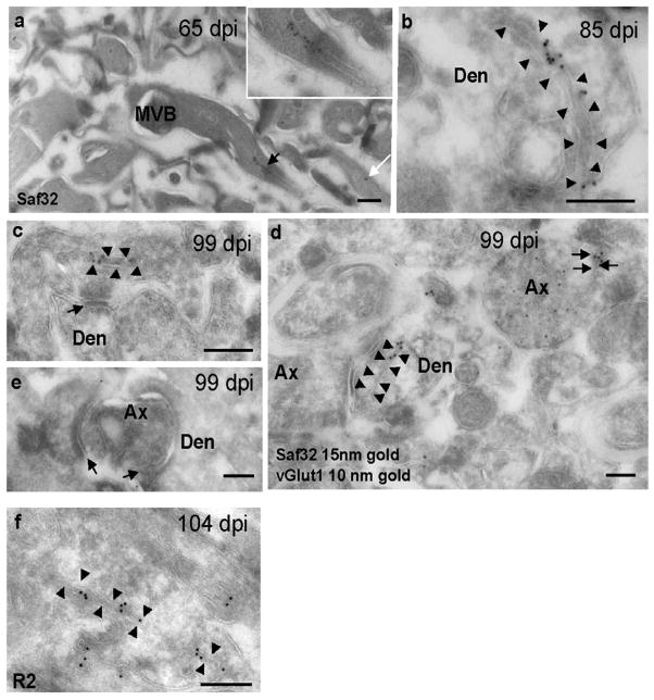
Figure 5. Cryo-immunogold EM labeling of PrPSc on membrane invaginations.
Hippocampal sections were taken from prion-infected mice at 65 dpi (a), 85 dpi (b), 99 dpi (c, d, e), 104 dpi (f). (a–d) Saf32 labeling of PrPSc on membrane invaginations in stratum oriens of prion-infected hippocampus. (a) Process with labeled paired membranes (black arrow) that continue to the surface membrane. The inset shows a higher magnification view of the process. The white arrow indicates another process with paired membranes labeled with two gold particles. (b) Dendrite, with labeled coated membranes (arrowheads) that extend to the plasma membrane. (c) Labeled paired membranes (arrowheads) in an axon terminal close to a synapse (arrow). (d) Saf32 labeling (15-nm gold) on a post-synaptic membrane invagination (arrowheads) and on paired membranes of an excitatory axon terminal (black arrows) co-labeled with anti-vGlut1 (10-nm gold). (e) Image showing degenerating axon terminal with no clustered Saf32 labeling on the axon or post-synaptic structures. Arrows indicate synapses. (f) Membrane invagination labeled with R2 antibody to the C-terminus of PrP. Abbreviations: Den, dendrite; Ax, axon; MVB, multivesicular body. Bars, 200 nm.

