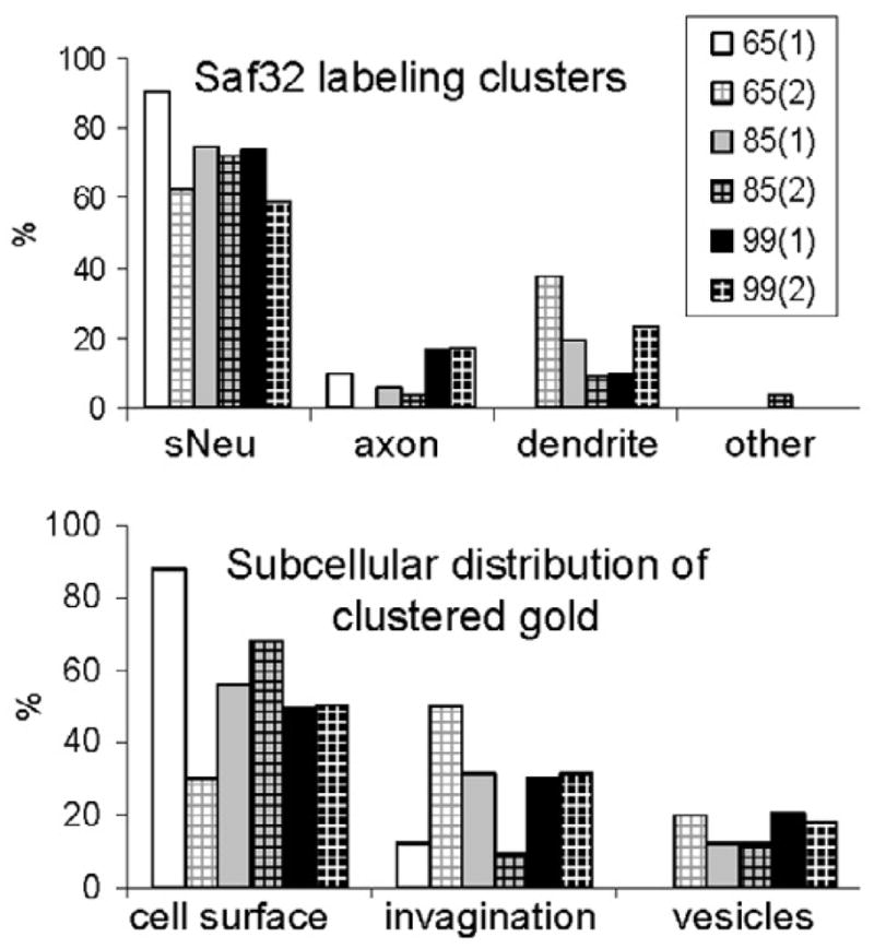
Figure 6. Quantification of clustered Saf32 labeling in infected hippocampus.

Quantification of Saf32 labeling, expressed as the proportional distribution of clustered gold particles in different types of neuronal structures (top) or subcellular compartments (bottom) at the timepoints indicated, in dpi. Numbers in parentheses indicate samples from different animals. “Other” indicates labeling in small vesicles of an astrocyte. Labeling on vesicles comprised 13% of the total clustered gold particles analyzed in infected hippocampus with 1.4% on synaptic vesicles. Abbreviations: sNeu, small neurite with diameter of <250 nm.
