
Fig. 1.
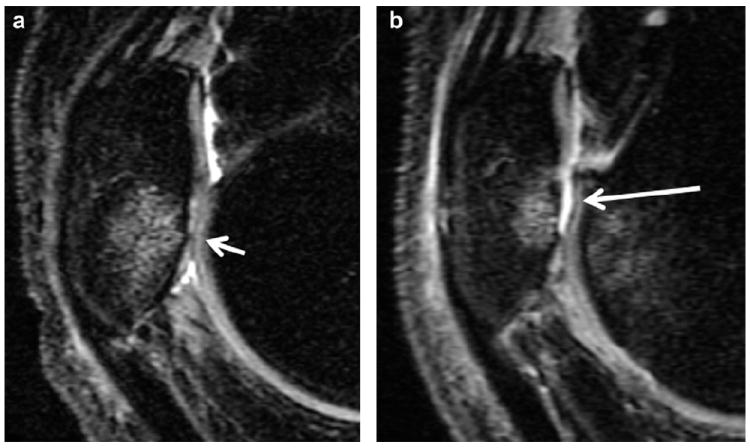
Sagittal, intermediate-weighted MR images demonstrate progression of a cartilage lesion at the PAT: at baseline (a) a small amount of partial-thickness cartilage loss at the inferior pole of the patella (short arrow) associated with bone marrow edema is seen and at the 12 month-follow-up (b) extensive full-thickness cartilage loss is shown at the same site (long arrow).