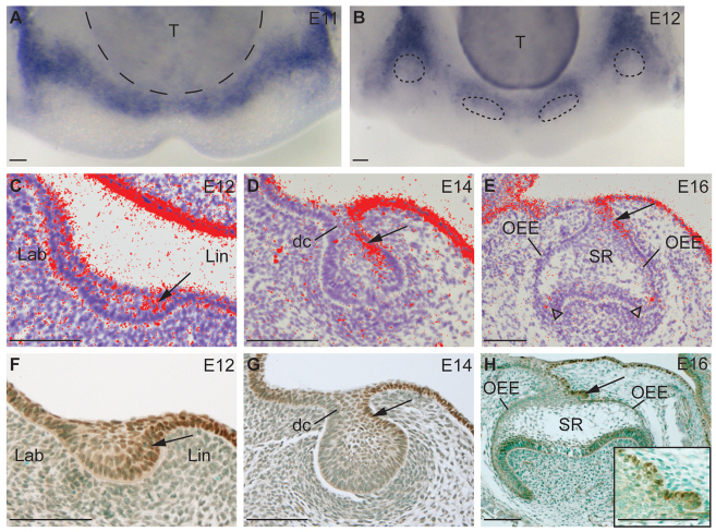
Fig. 2.
Sox2 is localized to primary dental lamina and to lingual dental epithelium during mouse molar development. (A,B) Whole-mount in situ hybridization showing expression of Sox2 mRNA (purple) in mouse lower jaw at E11 in the primary dental lamina (A), and at E12 in oral epithelium and lingual to the tooth placodes (B, dashed circles). (C-H) The expression of Sox2 mRNA (red, C-E) and protein (brown, F-H) in the lower molar from E12 to E16 is gradually restricted to lingual dental epithelium (arrows). Arrowheads in E point to Sox2 expression in E16 cervical loops. Arrow in H points to budding of dental epithelium at the lingual side of molar at E16 (inset shows higher magnification). dc, dental cord; Lab, labial; Lin, lingual; OEE, outer enamel epithelium; SR, stellate reticulum; T, tongue. Scale bars: 100 μm.

