
Fig. 5.
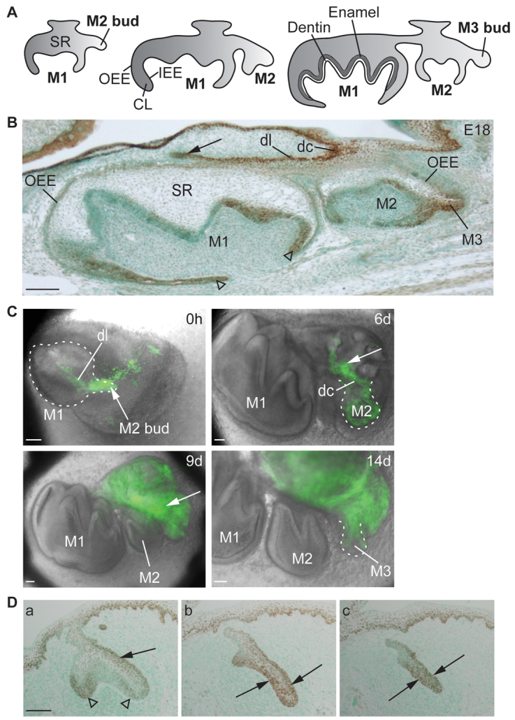
Sox2 expression is associated with successional formation of posterior molars in mouse and ferret. (A) Schematic of successional development of molars (see text for details). (B) Localization of Sox2 protein (brown) in E18 mouse molars. Sox2 is expressed in the dental cord and dental lamina connecting M1 to M2, and in the rudimentary dental lamina bud above M1 (arrow). Sox2 is expressed in the bud, which will form M3. Arrowheads point to Sox2 expression in M1 cervical loops. (C) Dynamics of Sox2 expression (green) during successional addition of molars. A dissected E14.5 M1 of a Sox2-GFP reporter mouse gives rise to M2 and M3 during 14 days of culture. The buds of M2 and M3 express Sox2-GFP but the completed crowns of M1 after 6 days (6d) and M2 after 9 days (9d) do not express Sox2-GFP. Arrows point to the GFP+ dental lamina, which will give rise to a new tooth. Dashed lines outline the tooth germs. (D) Localization of Sox2 protein (brown) in ferret M1 at E34 in frontal sections from anterior (a) to posterior (c). The planes of sections are shown by dashed lines in Fig. 3B. Sox2 localizes to the lingual OEE (a, arrow) and cervical loops of M1 (a, arrowheads). In the posterior end of M1 from where M2 develops, Sox2 localizes both to lingual and labial sides of M1 epithelium (b,c). Lingual is towards the right. CL, cervical loop; dc, dental cord; dl, dental lamina; IEE, inner enamel epithelium; M, molar; OEE, outer enamel epithelium; SR, stellate reticulum. Scale bars: 100 μm.