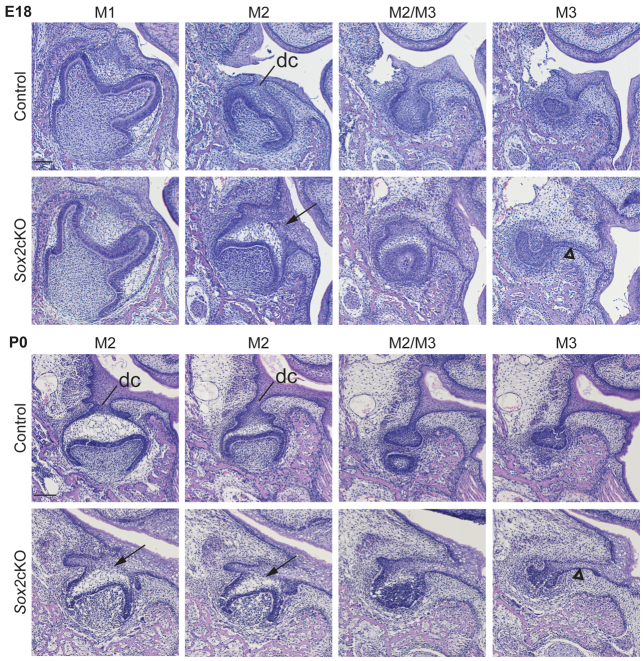
Fig. 7.
Conditional deletion of Sox2 leads to hyperplastic dental epithelium of M2 and M3. Hematoxylin and Eosin-stained serial frontal sections of mandibular molars of control and Sox2cKO mice at E18 and P0. Sox2cKO shows no obvious phenotype in M1 (data for P0 not shown), whereas in M2 the dental cord is expanded (arrows) and dental lamina between M2 and M3 is expanded. M3 is attached to oral epithelium by an extended sheet of dental lamina (arrowheads). See also supplementary material Fig. S2. dc, dental cord. Lingual is towards the right. Scale bars: 100 μm.

