
Fig. 8.
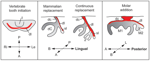
Sox2 expression is associated with epithelial competence of dental lamina in different modes of successional tooth formation. Schematic of the localization of Sox2 (red) in the primary dental lamina in the lower jaw, and in the dental lamina during different types of successional tooth formation: mammalian tooth replacement, continuous replacement and molar addition. The drawings also illustrate the morphological similarity between the two modes of successional tooth formation. 1°, first generation tooth; 2°, second generation tooth; A, anterior; B, buccal; dc, dental cord; dC, deciduous canine; dl, dental lamina; L, lingual; Le, left; M, molar; P, posterior; Ri, right; sl, successional dental lamina; t, tongue.