
Fig. 4.
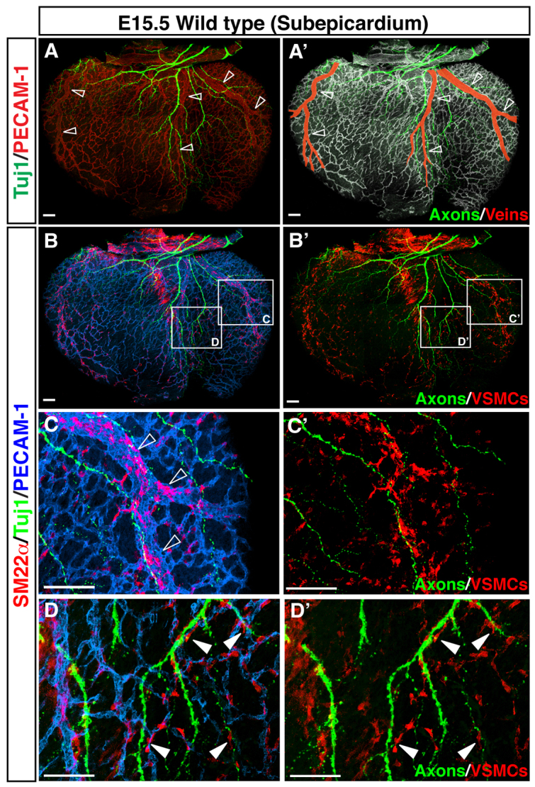
VSMCs cover nerve-associated large diameter coronary veins. (A-D′) Visualization of coronary VSMCs in the subepicardium. Whole-mount triple immunofluorescence confocal microscopy was performed with antibodies to the VSMC marker SM22α (B-D′, red) in addition to PECAM1 (A, red; A′, white; B-D, blue) and TUJ1 (green). Magnified images (C-D′) show the boxed regions in B,B′. At E15.5, SM22α+ VSMCs are found predominantly around nerve-associated large diameter coronary veins (A-C′, open arrowheads), and a smaller number of SM22α+ VSMCs are located between remodeled veins (D,D′, arrowheads). Scale bars: 100 μm.