
Fig. 2.
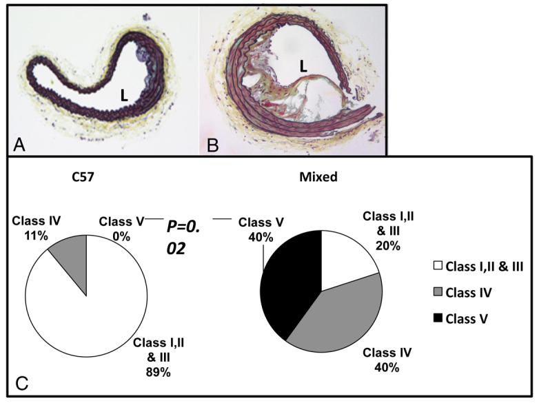
Comparison of brachiocephalic lesions in 16-week-old stressed mice of different backgrounds. (A,B) Brachiocephalic lesions of 16-week-old stressed C57 (A) and stressed mixed-background (B) mice. (C) Pie chart shows distribution of different lesion grades between C57 and mixed-background mice.