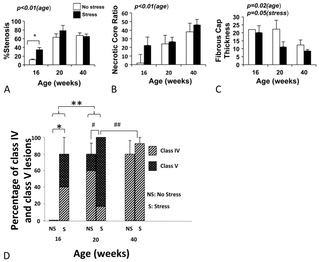
Fig. 4.
Percentage stenosis, ratio of lipid core to lesion area, fibrous cap thickness and percentage of class IV and class V lesions in stressed and unstressed mice of different ages. (A) Percentage stenosis increases with age from 16 to 20 weeks (P<0.01). At 16 weeks, stressed animals had greater percentage stenosis than unstressed animals (*P=0.02). (B) Ratio of lipid core to lesion area increases with age (P<0.01). (C) Fibrous cap thickness decreases with age (P=0.02). There is a trend for thinner fibrous cap in stressed animals (P=0.05). At 16 weeks, only one non-stressed mouse had a fibrous cap. (D) Percentage of class IV and V lesions increases with stress in 16-week-old mice (*P=0.02) and with age from 16 to 20 weeks (**P=0.01). Complex atheroma (class V) increases significantly with stress at the age of 20 weeks (#, P=0.03); however, there are significantly less complex atheroma (class V) in the 40-week-old mice compared with 20-week-old mice (##, P<0.01). All stressed animals at 20 weeks (100%) had class IV or V lesions.

