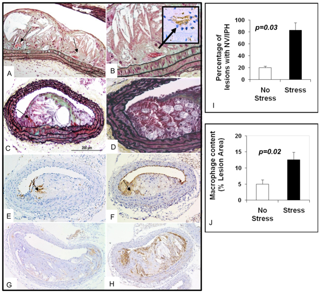
Fig. 5.
Neovascularization, intraplaque hemorrhage and inflammation in brachiocephalic lesions of a 20-week-old mixed-background ApoE−/− stressed mouse. (A) Longitudinal section of a 20-week-old brachiocephalic lesion shows neovascularization (arrows). (B) Same lesion as in A with higher magnification; CD 31 staining confirms the presence of neovascularization (arrow). (C) Cross-section of a lesion with Movat staining shows RBCs (arrow) in the plaque. (D) Movat staining of a different cut of the same lesion with higher magnification shows the presence of intraplaque hemorrhage in the shoulder area. (E)Ter119 staining of the same lesion confirms the presence of RBCs (arrow). (F) CD 31 staining of the same lesion shows presence of endothelial cells (arrow) in the plaque. (G,H) Mac3 staining of 20-week-old mixed-background ApoE−/− non-stressed (G) and stressed mice (H). (I) Bar graphs show that 20-week-old mixed-background ApoE−/− stressed mice have more neovascularization (NV) and/or intraplaque hemorrhage (IPH) than non-stressed mice. (J) Bar graph shows greater degree of macrophage content in lesions from 20-week-old mixed-background ApoE−/− stressed animals compared with non-stressed mice of the same age.

