Fig. 2.
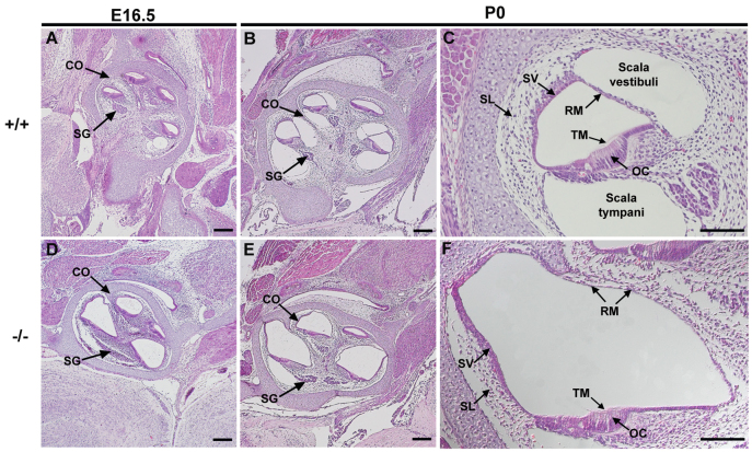
Cochlear duct of Atp6v0a4 mutants is severely enlarged. (A-F) H&E staining of mid-modiolar Atp6v0a4 inner ear at E16.5 (A,D) and P0 (B,C,E,F). At E16.5 and P0 homozygous mutants (D-F) show an enlargement of the entire cochlear duct compared with controls (A-C). A higher magnification of the organ of Corti (C,F) shows the abnormal expansion of the scala media. Reissner's membrane is distended towards the scala vestibuli and the tectorial membrane is observed uncovered by any cellular layer (F), indicating that the membrane is not collapsed or fused to the organ of Corti. CO, cochlea; SG, spiral ganglion; SL, spiral ligament; SV, stria vascularis; OC, organ of Corti; RM, Reissner's membrane; TM, tectorial membrane. Scale bars: 200 μm (A,B,D,E); 100 μm (C,F).

