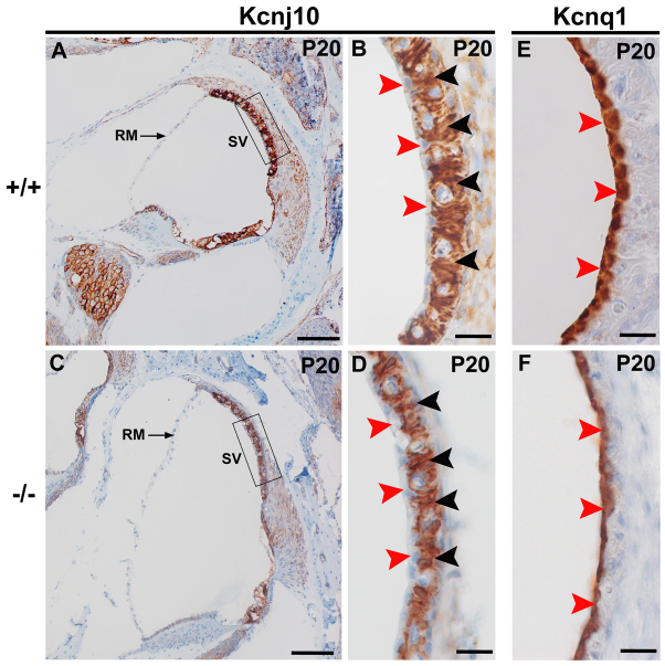
Fig. 6.
Kcnj10 and Kcnq1 are expressed in the stria vascularis of Atp6v0a4 mutant mice. (A,C) Mid-modiolar sections of the inner ear of control (A) and mutant (C) mice at P20. Regions in boxes in A and C are magnified in B and D, respectively. In wild types and mutants, a similar Kcnj10 expression pattern is observed. Especially strong expression is found in the intermediate cells of the stria vascularis (B,D, black arrowheads), whereas the marginal cells are negative (B,D, red arrowheads). It is also noticeable that mutants examined (n=5) did not seem to show Reissner's membrane so much distended towards the scala vestibuli as at P0 and P5. We think this might be due to the decalcification process used to facilitate the sectioning of these samples, or to the resolution of any pressure difference across Reissner's membrane by rupture. (E,F) Expression of Kcnq1 is specifically found in the marginal cells of the stria vascularis at P20 in both wild types and mutants (E,F, red arrowheads). RM, Reissner's membrane; SV, stria vascularis. Scale bars: 100 μm (A,C); 20 μm (B,D-F).

