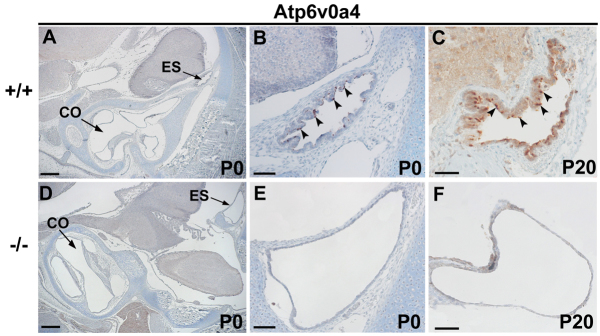
Fig. 7.

Expression of Atp6v0a4 in inner ear. (A-F) Sections of controls (A-C) and mutants (D-F) at P0 (A,B,D,E) and P20 (C,F) stained by immunohistochemistry. In wild-type mice at both ages, the expression of a4 subunit was specifically found in a subpopulation of cells within the ES epithelium (B,C, arrowheads). No expression was observed anywhere else within the inner ear (A). In Atp6v0a4 knockouts, no expression of this protein was detected (E,F). CO, cochlea. Scale bars: 300 μm (A,D); (B,E) 200 μm; 50 μm (C,F).
