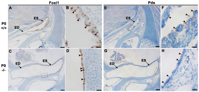
Fig. 8.
Foxi1 and Pds are expressed in the ES of Atp6v0a4 mutants. (A-D) Expression of Foxi1 in the endolymphatic duct and sac of wild-type and mutant mice at P0. (B,D) Higher magnification of the boxed regions in A and C, respectively. In control mice, Foxi1 is strongly expressed in a subpopulation of cells within the infolded epithelium of the ES (B, arrowheads). A similar pattern is observed in the flattened epithelium of the ES in mutants (D, arrowheads). (E-H) Expression of Pds in the ES of wild-type and mutant mice at P0. (F,H) Higher magnification of the boxed regions in E and G, respectively. In wild types, Pds expression is found in a few scattered cells within the ES (F, arrowheads). A similar pattern is found in the flattened ES seen in mutants (H, arrowheads). ED, endolymphatic duct. Scale bars: 100 μm (A,C,E,G); 20 μm (B,D,F,H).

