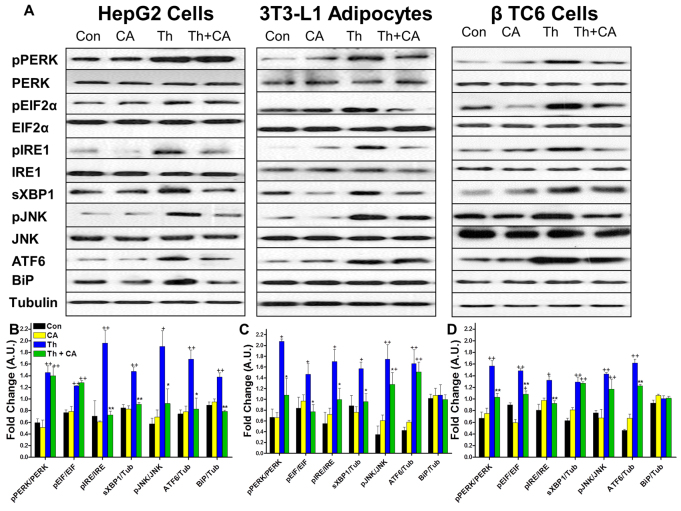
Fig. 8.
Preincubation of hepatocytes, adipocytes and β-cells with cholate protects cells from the development of ER stress. Cells were incubated with 100 μM cholic acid or DMSO for 24 hours then exposed to 2 μM thapsigargin for 2 hours. (A) Representative immunoblots for pPERK(Thr980) and total PERK, peIF-2α(Ser51) and total eIF2α, pIRE1(Ser724) and total IRE1, sXBP1, pJNK(Thr183/Tyr185) and total JNK and BiP. Experiments were performed in HepG2, differentiated 3T3 L1 adipocytes and β-TC6 cells. (B-D) Control cells (Con) were not exposed to cholic acid or thapsigargin. Other cells were exposed to chloic acid but not to thapsigargin (CA), to thapsigargin but not cholic acid (Th), or preincubated with cholic acid and then exposed to thapsigargin (Th + CA). Results were quantified in densitromic units and expressed relative to the total protein of interest or relative to tubulin for BiP, ATF6 and sXBP1 in hepatocytes (B), adipocytes (C) and β-cells (D). *P<0.05, **P<0.01 for IT compared with Th by Student's t-test. +P<0.05, ++P<0.01 compared with Con and CA by Student's t-test; n=3 per group.

