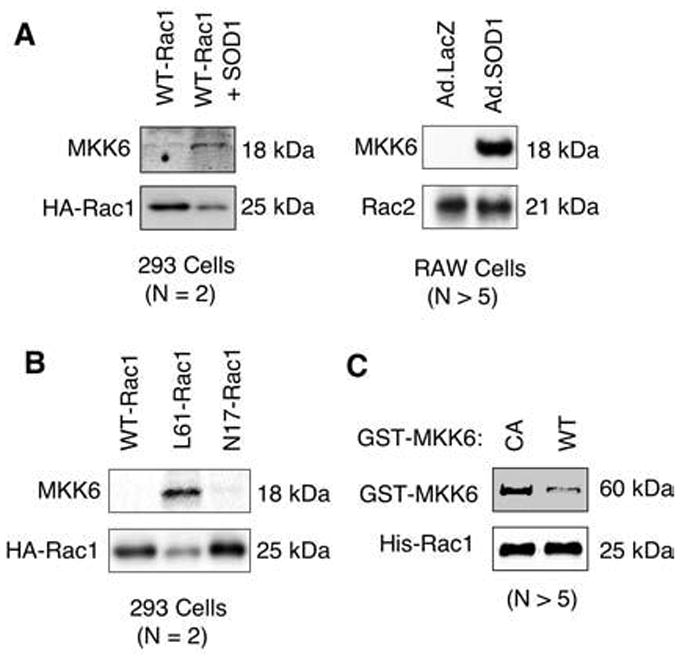
FIG. 2. The 18-kDa MKK6 fragment binds Rac in 293 cells and RAW cells under conditions that elevate cellular hydrogen peroxide.

(A, B) The 293 cells were transfected with the following plasmids expressing HA-tagged WT-Rac1, L61-Rac1, N17-Rac1, or WT-Rac1+SOD1. HA-Rac1 was then immunoprecipitated from the 293 cell lysates by using an anti-HA antibody, and the proteins that associated with Rac1 were separated by SDS-PAGE and characterized by Western blotting with the indicated antibodies. In a similar set of experiments, association of endogenous Rac2 with MKK6 was assessed in RAW cells infected with Ad.LacZ or Ad.SOD1. Cell lysate from each condition was immunoprecipitated with anti-Rac2 antibody and evaluated with Western blot. (C) In vitro association of purified GST-CA-MKK6 and GST-WT-MKK6 with His-tagged Rac1. His-tagged Rac1 was immobilized on Dynabeads, and pull-down assays were performed with an equal amount of purified CA or WT GST-MKK6. Proteins bound to Rac1 were evaluated with Western blot by using anti-GST and anti-Rac1 antibodies. All panels are representative for n = 2–5 independent experiments as indicated.
