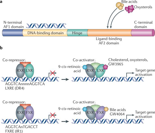
Figure 1. Mechanism of action of LXR and FXR.
a | The basic structure of a nuclear receptor, highlighting the DNA-binding and ligand-binding domains. b | Liver X receptor (LXR) forms an obligate heterodimer with retinoid X receptor (RXR) that binds to a DR4 (direct repeat spaced by four nucleotides) LXRE (LXR response element) in the regulatory regions of target genes, thereby repressing gene expression. Following ligand binding to LXR or RXR, the heterodimer changes conformation, which leads to the release of co-repressors and the recruitment of co-activators. This results in the transcription of target genes. Similarly, farnesoid X receptor (FXR) forms a heterodimer with RXR and binds to the FXR response element (FXRE), which is typically an inverse repeat spaced by one nucleotide (IR1), in its target genes to induce gene expression. AF domain, activation function domain; C-terminal, carboxy-terminal; N-terminal, amino-terminal.

