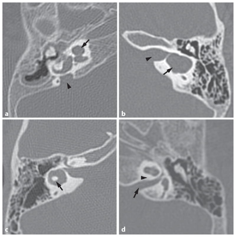
Fig. 1.

A variety of CT temporal bone images showing anomalies encountered in children with congenital hearing loss. a Axial CT of a right temporal bone with a Mondini malformation of the cochlea (arrow) and an enlarged vestibular aqueduct (arrowhead). b Axial CT of a left temporal bone with a common cavity malformation (arrow) and bony separation of common cavity from the IAC (arrowhead). c Axial CT of a right temporal bone with lateral semicircular canal dysplasia – the bone island in the center of the canal (arrow) is too small. d Axial CT of a left temporal bone with a narrow IAC (arrow) and no cochlear aperture for the auditory nerve to enter the cochlea (expected location is marked by arrowhead).
