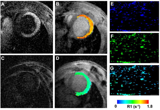
Figure 1.
Molecular MRI of cardiomyocyte necrosis with the DNA-binding agent Gd-TO. (A, B) Infarcted mouse imaged 2 hours after the injection of Gd-TO. (C, D) Infarcted mouse imaged 2 hours after the injection of Gd-DTPA. While Gd-DTPA has washed out of the infarct, Gd-TO has bound to the exposed DNA of the necrotic cells. This manifests as an increase in signal intensity on the T1 weighted image (A) and an increase in the longitudinal relaxation rate R1 (B). (C, D) No increases in signal intensity or R1 are seen in the mouse injected with Gd-DTPA. Fluorescence microscopy of the excised heart confirms the binding of Gd-TO to DNA in the infarct. (E) DAPI, (F) Gd-TO, (G) fused image. Reproduced with permission [6].

