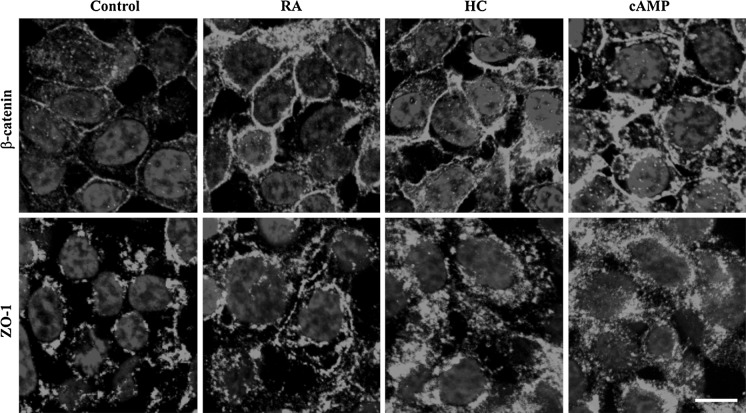
Fig. 2.
Immunohistochemical staining of RPMI 2650 cells for junctional proteins β-catenin and zona occludens-1 (ZO-1) visualized by confocal fluorescence microscopy. Cells were treated with retinoic acid (RA, 300 μg/mL), hydrocortisone (HC, 500 nM) or 3′–5′-cyclic adenosine monophosphate (cAMP, 250 μM) for 24 h; bar 10 μm. Control conditions: RPMI 2650 cells were grown in Eagle’s minimal essential medium supplemented with 10 % foetal bovine serum and 50 μg/mL gentamicin

