
Fig. 1.
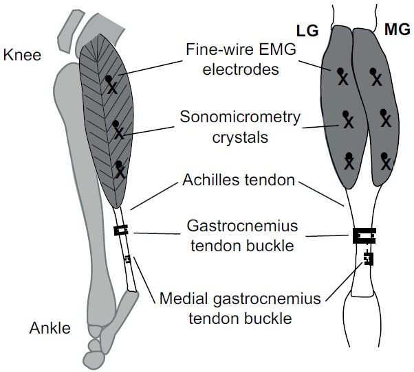
Lateral (left) and posterior (right) view of the goat lower hindlimb. The approximate locations of the electromyographic (EMG) electrodes and sonomicrometry crystals (proximal, mid-belly and distal) in the lateral and medial gastrocnemius (LG and MG) muscles, and the common gastrocnemius and MG tendon-buckle force transducers are shown (adapted from Lee et al., 2011).