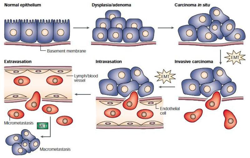
FIGURE 2.
The epithelial-mesenchymal transition and mesenchymal-epithelial transition (MET) model of tumor metastasis. According to Jean Paul Thiery, normal epithelia lined by a basement membrane can proliferate locally to give rise to an adenoma. Further transformation by epigenetic changes and genetic alterations leads to a carcinoma in situ, still outlined by an intact basement membrane. Further alterations can induce local dissemination of carcinoma cells, possibly through an EMT, as the basement membrane becomes fragmented. The invasive carcinoma cells (red) then intravasate into lymph or blood vessels, allowing their passive transport to distant organs. At secondary sites, solitary carcinoma cells extravasate, remain solitary (micrometastasis), or form a new carcinoma through an MET. Reprinted with permission from18.

