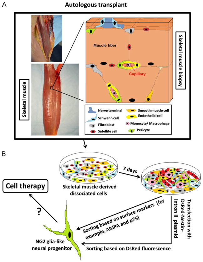
Fig. 8.
Diagram representing the origin of Nestin-GFP+ progenitor cells derived from a skeletal muscle biopsy. (A) The skeletal muscle consists of bundles of multinucleated myofibers, each carrying a population of satellite cells. Various mesenchymal cells are present in the muscle interstitium: fibroblasts (gray), smooth muscle cells (orange), endothelial cells (yellow), and pericytes (green), which emit processes along and around capillaries. The communication between the motor axon terminal (dark blue) and muscle fiber (neuromuscular junction) is represented. Schwann cells associated with the nerve terminal are shown (light blue). (B) Various cell types are represented in enzymatically dissociated muscles cultured for 7 days: fibroblasts (gray), smooth muscle cells (orange), endothelial cells (yellow), macrophages (pink) derived from circulating monocytes, Schwann cells (light blue), pericytes (green), multinucleated myotubes (red) and myoblasts (red) derived from satellite cells (red), and a small population of neural progenitor cells (NG2-glia like cells) (green) derived from GFP+ cells (green). Surface markers or use of DsRed-Nestin-Intron II plasmid (Fig. 2) allow neural progenitor cell sorting and eventual application for cell therapy. (For interpretation of the references to color in this figure legend, the reader is referred to the web version of this article.)

