Figure 5.

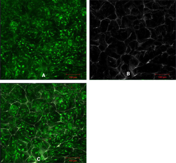
Confocal micrograph of a 3-D culture of HDF into a chitosan on day 14. Scale bar 100 μm. Live cell imaging of HDF (A). The unstained architecture of the chitosan (B). 3-D cultured cells into a chitosan scaffold (C).

Confocal micrograph of a 3-D culture of HDF into a chitosan on day 14. Scale bar 100 μm. Live cell imaging of HDF (A). The unstained architecture of the chitosan (B). 3-D cultured cells into a chitosan scaffold (C).