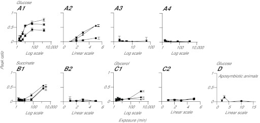
Fig. 4.

Detection of labeled glucose, but not of other compounds, in host tissue at early times after the exposure of symbiotic anemones to light and 13C-bicarbonate. (A–C) CC7 anemones were incubated for various times in the light or dark with 13C-bicarbonate or 12C-bicarbonate, as indicated, and were then homogenized and analyzed as described in the Materials and methods. The peak-ratio method was used to determine the distributions of 13C-labeled glucose (A), succinate (B) and glycerol (C) in the total homogenate (circles), the filtrate (triangles), and the material retained on the filter (squares). (1) Dark pre-incubation followed by incubation in the light with 13C-bicarbonate; (2) light pre-incubation for ~2 h followed by incubation in the light with 13C-bicarbonate; (3) dark pre-incubation followed by incubation in the dark with 13C-bicarbonate; (4) dark pre-incubation followed by incubation in the light with 12C-bicarbonate. (D) As in panel A1 except using aposymbiotic anemones. Time axes are in log or linear scales, as indicated. Error bars (all panels) represent 95% confidence intervals based on the three biological replicates in each case; some error bars in A2, B1 and C1 are offset from the corresponding data points to allow easier discrimination between the symbols.
