
Fig. 4.
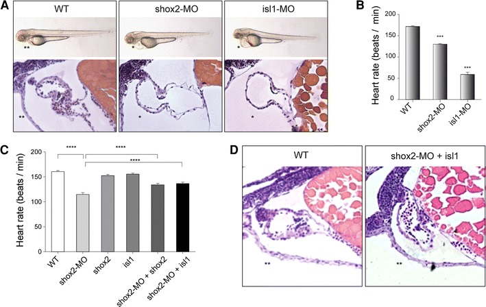
shox2-MO-mediated bradycardia can be rescued by isl1 overexpression. A Injection of 2.8 ng shox2 and isl1 antisense morpholinos (shox2-MO and isl1-MO) leads to pericardial edema and pericardial blood congestion due to reduced heart rates in zebrafish embryos. Lateral views of WT, shox2-MO and isl1-MO injected embryos 72 h post fertilization (hpf). Hematoxylin and eosin staining of sagittal histological sections of WT (left), shox2-MO (middle) and isl1-MO (right) morphant hearts 72 hpf. Endocardial and myocardial layers of ventricle and atrium are defined, and heart chambers are separated by an atrioventricular ring. B Heart rates of shox2 and isl1 morphants are decreased by 72 hpf. Injection of shox2-MO leads to sinus bradycardia with heart frequencies of 130 beats per min (bpm) compared to WT embryos with 172 bpm. isl1 deficient embryos exhibit severe sinus bradycardia and frequent pauses in the cardiac contraction with heart frequencies of 59 bmp. (***P < 0.001). C shox2-MO-mediated bradycardia is partially rescued by cardiomyocyte-specific expression of isl1. The mean heart rate of WT embryos 72 hpf amounts to 160 bpm. After shox2 knockdown (2.8 ng shox2-MO) the heart rate is reduced to 114 bpm. Cardiomyocyte-specific overexpression of shox2 or isl1 alone (0.32 ng) does not affect the heart beat. Coinjection of 0.32 ng shox2 or isl1 plasmid DNA along with 2.8 ng shox2-MO results in significantly increased heart frequencies (134 and 137 bmp) compared to shox2-MO-injected embryos (114 bpm) at 72 hpf (****P < 0.001; n = 32–40 per condition). D Hematoxylin and eosin staining of sagittal histological heart sections of WT (left) and shox2-MO embryos rescued by isl1 overexpression (right) 72 hpf revealed that the rescued hearts, compared to those of shox2-MO injected embryos (A, middle picture), show a phenotype very similar to the WT (left). WT wildtype, asterisk pericardial edema, double asterisks no pericardial edema