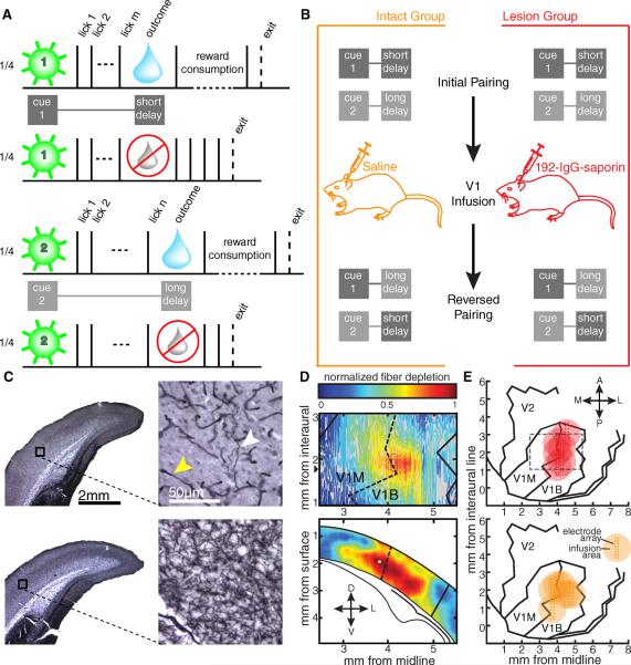
Fig 1.
Experimental design. A) Following a 2 s intertrial interval, rats could enter the nosepoke to activate one of four pseudorandomly interleaved trial types. A brief cue (green flash) was presented to either the left eye or the right and was predictive of the number of licks (solid black ticks) required to gain a small bolus of water (blue drop) in half of the trials. B) Implanted animals performed the task as outlined in A before receiving a bilateral infusion of either saline or 192-IgG-saporin into V1. Following recovery, the task parameters were reversed such that the cue previously associated with the short delay was now paired with the longer delay and vice versa. C) Coronal sections demonstrating AChE histochemistry from a lesioned animal (top) and an intact animal (bottom). The black boxes on the low magnification images (left) correspond to the high magnification regions on the right. The white arrowhead indicates a remaining fiber stained for AChE, and the yellow arrowhead indicates a capillary. D) Example visualization of fiber depletion. The contour plot (top) shows the relative difference of laminar-averaged staining intensity between atlas-matched sections from an intact and lesioned hemisphere. The heat plot (bottom) represents the relative comparison for the coronal slice with the widest apparent lesion extent (anterior-posterior position indicated by the black triangle next to the contour plot above). The solid lines demarcate the boundary of V1 while the dashed lines indicate the border between the monocular (V1M) and binocular (V1B) portions of V1. Lower diagram adapted from Paxinos and Watson (2008). E) Aerial view of approximate recording locations and infusion zones for the lesion (top) and intact (bottom) groups. Each large circle indicates the expected region affected by the infusion for each animal, and the estimated position of the 8×2 recording electrode array is indicated by the pin points. The gray dashed line indicates the region shown in the contour plot in D. Aerial maps adapted from Zilles (Zilles, 1985).

