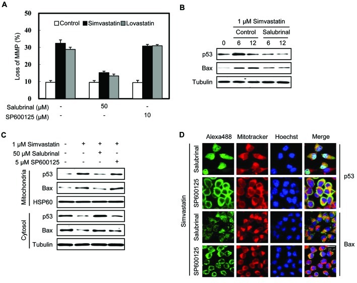
Figure 3.
Inhibition of eIF2α dephosphorylation by salubrinal decreases the stabilization and translocation of p53 to the mitochondria in simvastatin-induced apoptosis. (A) MethA cells pre-incubated with 50 μM salubrinal or 10 μM SP600125 for 30 min were treated with 1 μM simvastatin or lovastatin for 16 h. The cells were then stained with rhodamine-123 and relative fluorescence intensity was analyzed by flow cytometry to measure the loss of MMP. (B) MethA cells were pre-incubated with 50 μM salubrinal for 30 min and were then treated with 1 μM simvastatin for the indicated time periods. The level of p53 and Bax proteins were assessed by western blot analysis. (C) MethA cells were pre-incubated with 50 μM salubrinal or 5 μM SP600125 for 30 min and then treated with 1 μM simvastatin for 12 h. The cells were divided into cytosolic and mitochondrial fractions and the levels of p53 and Bax in the cytosolic and mitochondrial fractions were assessed by western blot analysis. (D) MethA cells were incubated under the indicated conditions for 12 h and immunostained with anti-p53 and anti-Bax antibody (green). p53 and Bax were observed under a confocal microscope at ×630 magnification. Nuclear staining with Hoechst dye (blue) and mitochondrial staining with MitoTracker (red) are shown.

