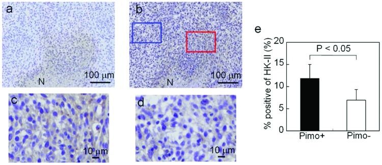
Figure 6.
HK-II expression in comparison with pimonidazole distribution. Representative images of (a) pimonidazole and (b) HK-II IHC staining. Typical immunostaining of pimonidazole in (c) Pimo+ and (d) Pimo−. (e) HK-II expression assessed by semiquantitative analysis in Pimo+ and Pimo−. N, necrotic/apoptotic regions.

