
Figure 4.
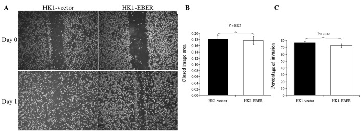
Scratch-wound assay and in vitro FluoroBlok tumour invasion assay. (A) A line scratch was made and photomicrographs were captured at days 0 and 1 (magnification, ×50). (B) Using the TScratch program, a software tool for automated analysis of wound healing assays, the open image area was calculated; from which the closed imge area (reflecting the degree of migration or would healing) was derived. No significant difference in migration ability was identified between the two stable cell lines. (C) Quantification of fluorescence emitted by cells that have invaded to the underside of the FluoroBlok membrane was detected with a plate reader and the % of cell invasion was calculated. No significant difference in invasiveness was observed between the cell lines. EBERs, Epstein-Barr virus-encoded RNAs.