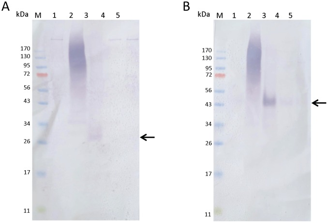
Figure 4. Expression of PpPer1 and PpPer3 in P. papatasi midgut lysates.
Expression of native PpPer1 and PpPer3 in P. papatasi midgut lysates was assessed using one midgut equivalent (from pools of five guts for each time point) of non blood fed and blood fed P. papatasi per lane. Lanes: M, size marker; 1, non blood fed midgut; 2, blood fed midgut dissected 24 h PBM; 3, blood fed midgut dissected 48 h PBM; 4, blood fed midgut dissected 72 h PBM; 5, blood fed midgut dissected 96 h PBM. Proteins were transferred to nitrocellulose and blots were incubated with anti-PpPer1 (A) and with anti-PpPer3 (B) specific antisera. Arrows point the native proteins PpPer1 (A) and PpPer3 (B).

