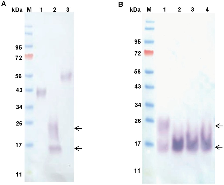
Figure 6. Recombinant proteins.
Western blots were carried out with 6×His-tagged rPpPer1, rPpPer2, and rPpPer3 (250 ng each protein) obtained from FreeStyle CHO-S cells. Proteins were separated on 4–12% reducing NuPAGE gels. (A) Proteins were transferred to nitrocellulose and incubated with anti-His antibody (1∶2,000), followed by anti-mouse AP-conjugated (1∶10,000). Lanes: M, molecular weight marker; 1, PpPer1; 2, PpPer2; 3, PpPer3. (B) rPpPer2 (200 ng) was N-deglycosylated with 1,000 (lane 2), 2,000 (lane 3) and 3,000 (lane 4) units of PNGase F for 16 h at 37°C (untreated rPpPer2 control lane 1). Separated proteins were transferred to nitrocellulose and incubated with anti-His followed anti-mouse AP-conjugated. M: molecular weight marker. Arrows indicate the 16 kDa and 20-to-24 kDa bands seen on rPpPer2 preparations (A and B). After PNGase F treatment the 20-to-24 kDa bands are no longer detected.

