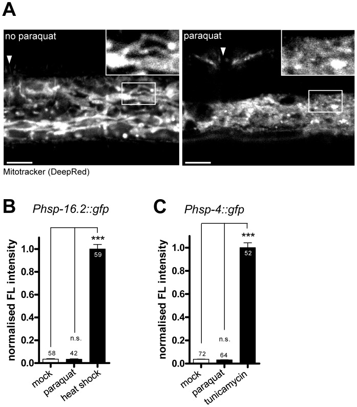
Figure 4. Paraquat affects mitochondrial morphology, but does not provoke the unfolded protein responses of the endoplasmatic reticulum (ER) or the cytosol.
A. Representative confocal micrographs of cells in wild type worms after Mitotracker staining. Worms were exposed to 0.5 mM paraquat starting from early L3 stage. Hypodermal mitochondrial staining was analyzed after two days. Arrows indicates the location of vulva. Scale bar: 10 µm. B–C. Quantification of GFP fluorescence intensities in a cytosolic UPR reporter strain (Phsp-16.2::gfp) (B) and an UPRER reporter strain (Phsp-4::gfp) after two days of exposure to 0.5 mM paraquat or to their respective inductor (heat shock: 4 h at 34°C at L4, analyzed after one day; tunicamycin: 7.2 µM at L1, analyzed after three days). Both UPR reporters were not induced by paraquat, suggesting that the compound does not affect protein folding environment in cytosol or ER, respectively. Columns represent normalized pooled values of three independent experiments plus standard error of the mean (SEM). Numbers in or on columns indicate the number of analyzed animals (cytosolic UPR ntotal = 159; UPRER ntotal = 188). ***: p<0.001, n.s: p>0.05; Kruskal-Wallis test plus Dunn's Multiple Comparison Test.

