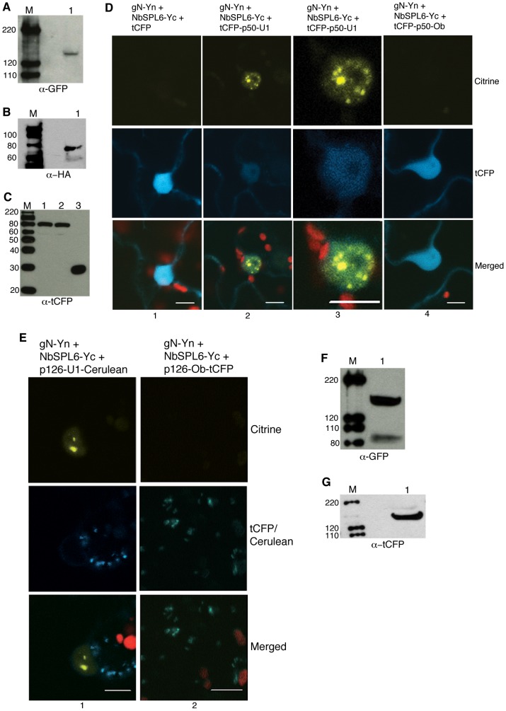
Figure 2. N associates with NbSPL6 in subnuclear bodies only during an active immune response.
A–C. Western blots showing gN-Yn (A), NbSPL6-HA-Yc (B), tCFP-p50-U1 (C, lane 1), p50-Ob-tCFP (C, lane 2), and tCFP (C, lane 3). M indicates marker. Protein sizes marked on the left are in kD. D. Co-expression of gN-Yn and NbSPL6-Yc with tCFP did not reconstitute citrine fluorescence (column 1) in BiFC assays. However, co-expression of gN-Yn and NbSPL6-Yc with tCFP-p50-U1 resulted in the reconstitution of citrine fluorescence within subnuclear bodies (column 2 and 3). Images in the column 3 are magnified versions of the nucleus shown in column 2. Citrine fluorescence was not observed when gN-Yn and NbSPL6-Yc were co-expressed with the non-eliciting p50-Ob-tCFP (column 4). Scale bars = 10 µm. The red structures are chloroplasts. E. Co-expression of gN-Yn and NbSPL6-Yc in the presence of the full-length 126 kD TMV-U1 replicase (p126-U1-Cerulean) reconstituted citrine fluorescence (column 1). Citrine fluorescence was not observed in the presence of the non-eliciting 126 kD replicase from the TMV-Ob strain (p126-Ob-tagCFP) (column 2). Scale bar = 10 µm. The red structures are chloroplasts. F and G. Western blots showing p126-U1-Cerulean (F) and p126-Ob-tCFP (G). M indicates marker. Protein sizes marked on the left are in kD.

