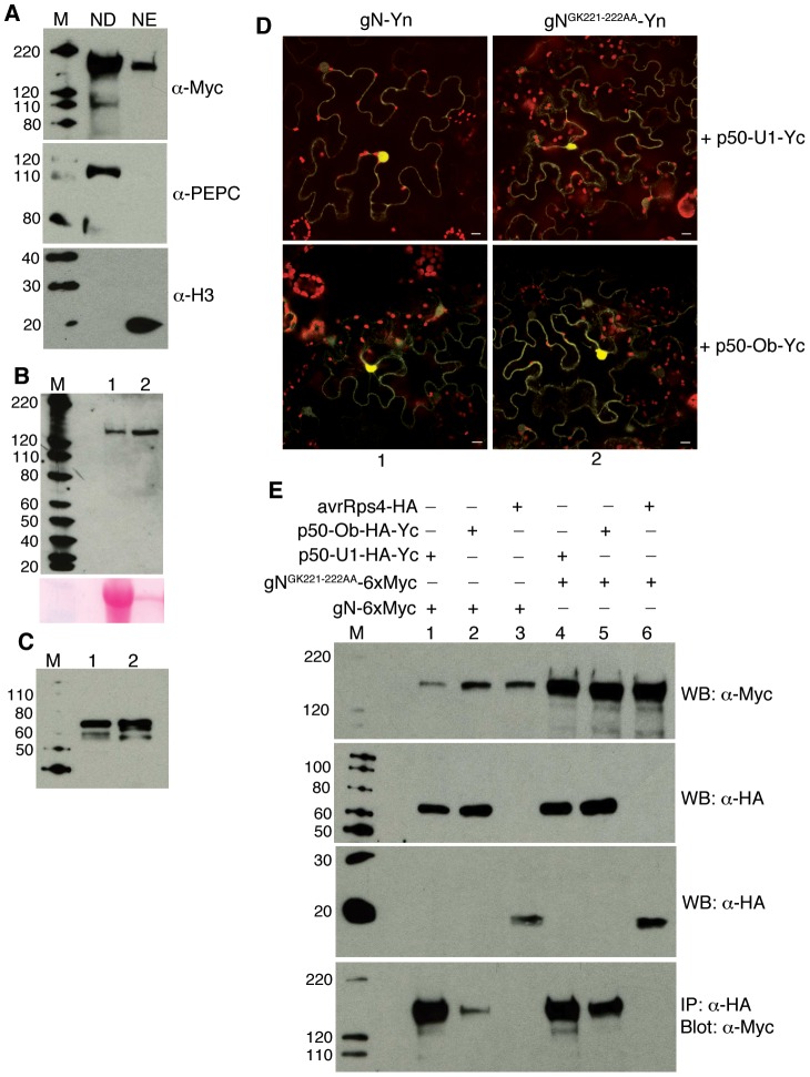
Figure 4. A P-loop mutant in the NB domain of the N immune receptor can still associate with p50-U1 or p50-Ob.
A. Cellular fractionation of gNGK221-222AA-TAP expressing tissue shows that the mutant protein is present in both the cytoplasmic fraction (ND) as well as the nuclear enriched (NE) fraction (upper panel). PEPC was used as a cytoplasmic marker (middle panel) and Histone 3 (H3) was used as a nuclear marker (bottom panel). M indicates marker. Protein sizes marked on the left are in kD. B–C. Western blots showing the expression of gN-Yn (B, upper panel, lane 1), gNGK221-222AA-Yn (B, upper panel, lane 2), p50-U1-HA-Yc (C, lane 1) and p50-Ob-HA-Yc (C, lane 2). The input volume for NGK221-222AA-Yn (B, upper panel, lane 2) was adjusted to 1/20th the volume loaded in lane 1 for gN (B, upper panel, lane 1). Ponceau staining (B, bottom panel) shows loading volume. M indicates marker. Protein sizes marked on the left are in kD. D. Co-expression of gN-Yn (column 1) or NGK221-222AA-Yn (column 2) with p50-U1- Yc (upper panels) and p50-Ob- Yc (lower panels) reconstitutes citrine fluorescence in BiFC assays. Scale bars = 10 µm. The red structures are chloroplasts. E. Co-immunoprecipitation of gN-6xMyc or gNGK221-222AA-6xMyc with p50-U1-HA-Yc, p50-Ob-HA-Yc or avrRps4-HA. Western blot analysis confirming expression of input proteins gN-6xMyc (panel1, lanes 1,2,3) and gNGK221-222AA-6xMyc (panel 1, lanes 4,5,6), p50-U1-HA-Yc (panel 2, lanes 1 and 4) and p50-Ob-HA-Yc (panel 2, lanes 2 and 5), and avrRps4-HA (panel 3, lanes 3 and 6). gN-6xMyc (panel 4, lanes 1 and 2) and gNGK221-222AA-6xMyc (lanes 4 and 5) co-immunoprecipitated with p50-U1 and p50-Ob. gN-6 myc or gNGK221-222AA-6xMyc did not co-immunoprecipitate with avrRps4 (panel 4, lanes 3 and 6). M indicates marker. Protein sizes marked on the left are in kD.

