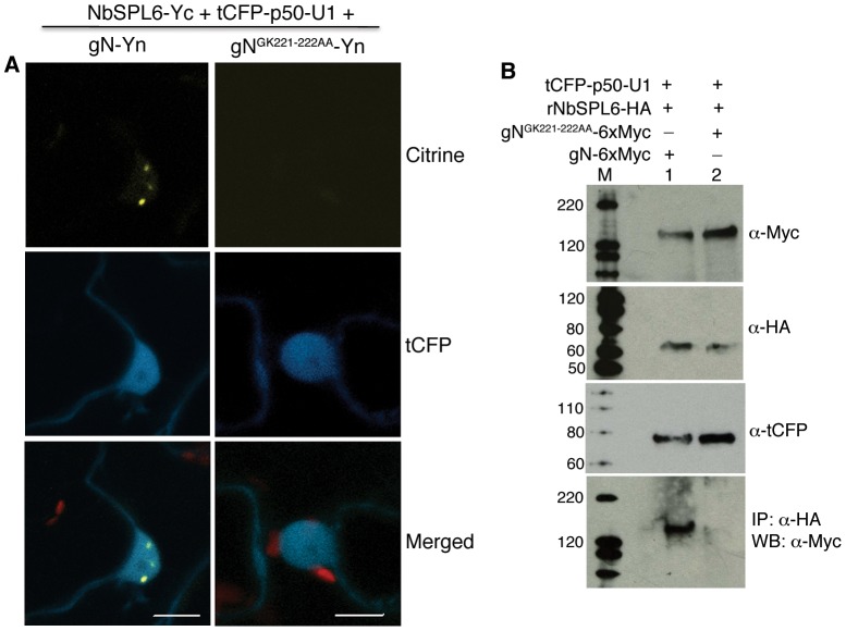
Figure 5. Mutation in the P-loop of the N immune receptor abolishes its association with NbSPL6.
A. BiFC assay showing that gN-Yn when co-expressed with NbSPL6-Yc reconstitutes citrine fluorescence in the presence of the defense eliciting tCFP-p50-U1 effector (left columns). gNGK221-222AA-Yn when co-expressed with NbSPL6-Yc fails to reconstitute citrine fluorescence in the presence of tCFP-p50-U1 (right columns). Scale bar = 10 µm. The red structures are chloroplasts. B. gNGK221-222AA-6xMyc is unable to co-immunoprecipitate with rNbSPL6-HA in the presence of tCFP-p50-U1. Western blot analysis confirmed expression of input proteins gN-6xMyc and gNGK221-222AA-6xMyc (panel 1), rNbSPL6-HA (panel 2), and tCFP-p50-U1 (panel 3). While gN-6Myc co-immunoprecipitated with rNbSPL6 in the presence of p50-U1 (panel 4, lane 1), gNGK221-222AA-6xMyc failed to co-immunoprecipitate with rNbSPL6 (panel 4, lane 2). M indicates marker. Protein sizes marked on the left are in kD.

