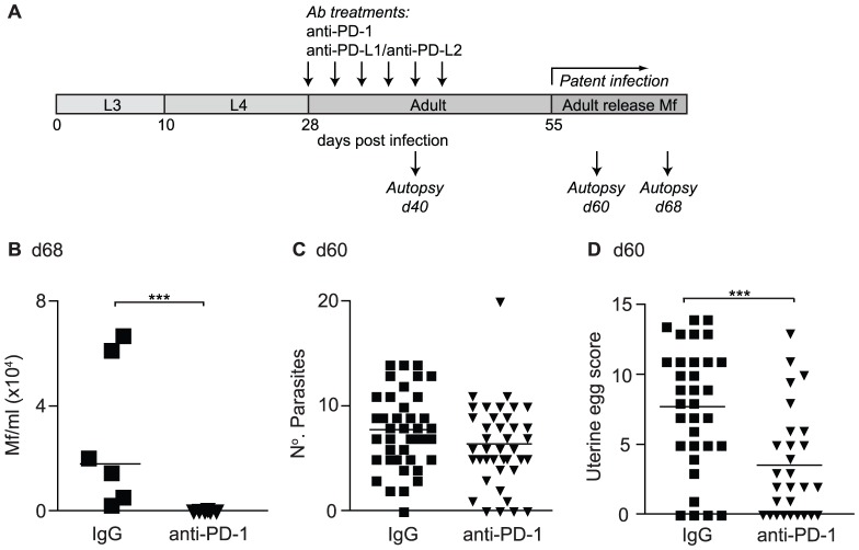
Figure 4. In vivo PD-1 blockade increases resistance to L. sigmodontis.
(A) Timeline of L. sigmodontis infection showing approximate timings of the molts from larval (L3/L4) to adult stages and the development of patency in relation to in vivo antibody treatments and autopsies. (B–D) L. sigmodontis-infected BALB/c IL-4gfp reporter mice were treated with a blocking anti-PD-1 mAb (triangles) or rat IgG (squares) from d28–d43 and their adult parasite burdens assessed at d 60 pi, and their blood Mf levels at d 68 pi. (B) Mf counts per ml of peripheral blood. (C) Number of adult parasites within the PC. (D) Number of live eggs within the uteri of individual female parasites recovered from IgG and anti-PD-1 treated hosts. Panels show one representative experiment of two (B) or pooled data from four independent experiments (C & D). Symbols represent individual mice (B & C) or female parasites (D), and lines represent means (B & C) or medians (D). *** p<0.001 (ANOVA performed on combined data from two (B) or four (C & D) independent experiments).

