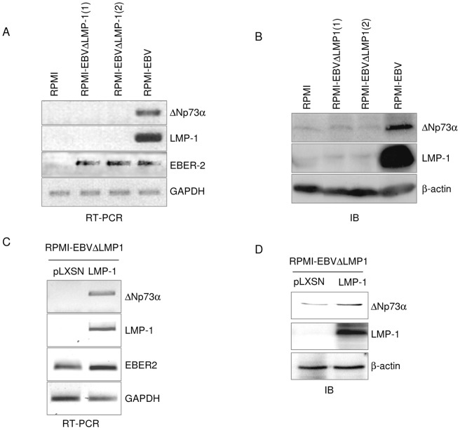
Figure 3. Expression of LMP-1 in cells infected by EBVΔLMP-1 restores ΔNp73α levels.
(A and B) Two independent infections of RPMI with EBVΔLMP-1 recombinant virus, not infected RPMI and RPMI carrying the wild-type EBV genome were collected and processed to obtain total RNA or whole cell extracts. (A) The levels of ΔNp73, LMP-1, EBER-2 and GAPDH transcripts were determined by RT-PCR. (B) Protein extracts were analyzed by immunoblotting using the indicated antibodies. Please note that lane 1 to 3 and lane 4 have been joined from 2 different areas of the same immunoblot. (C and D) Total RNA and total cell extracts from RPMI- EBVΔLMP-1 were transduced with pLXSN or pLXSN-LMP-1 retroviruses were prepared. (C) Transcript levels of ΔNp73α, LMP-1, EBER-2 and GAPDH were measured by RT-PCR. (D) Protein extracts were analyzed by immunoblotting for the levels of ΔNp73, LMP-1. β-actin was used as loading control.

