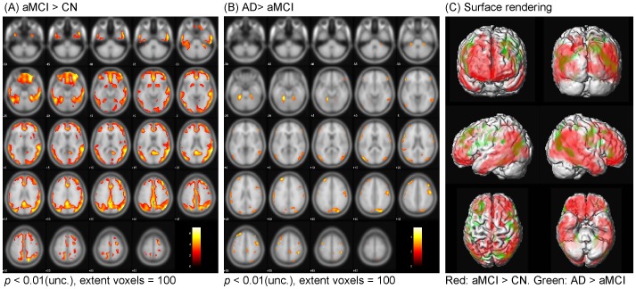
Figure 3. Statistical parametric mapping analysis: Localization of increased [18F]AV-45 retention between CN, aMCI and AD subjects.
Comparisons of [18F]AV-45 SUVRs between cognitively normal (CN) and amnestic mild cognitively impairment (aMCI) subjects (A), and between aMCI and Alzheimer's disease (AD) subjects (B) (Uncorrected for multiple comparisons and the color bar values indicate the value of the T-statistic in each display). Surface rendering was used to illustrate the cortical areas where [18F]AV-45 SUVRs were increased in aMCI than CN subjects (red) and increased in AD than aMCI subjects (green) (C).

