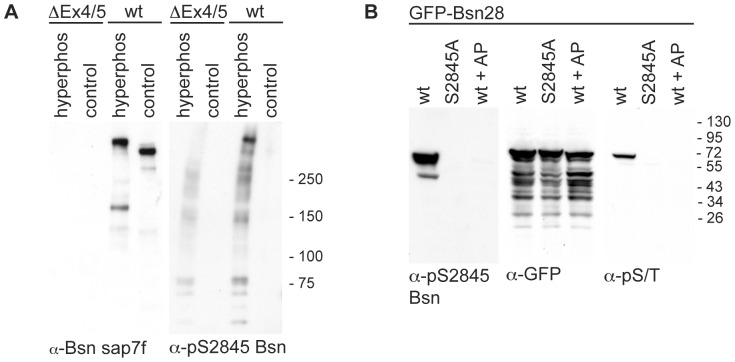
Figure 5. Characterization of the antibody against phosphorylated S2845 of Bassoon.
(A) α-pS2845 Bsn antibody was tested on P2 fractions from brains of wild-type (wt) and Bassoon mutant (BsnΔEx4/5) mice. Bassoon was detected by α-Bsn sap7f antibody in the samples from wt mice but not from mutant. The Western blot, which was prepared in parallel and incubated with the phosphorylation-specific antibody α-pS2845 Bsn preferentially detects Bassoon in the hyperphosphorylated sample and showed only weak unspecific immunoreactivity in the samples of the Bassoon-mutant mice. Images shown are representative for independent experiments done with lysates obtained from three pairs of mice. (B) GFP-Bsn28 (wt) or its mutant (S2845A) were expressed in HEK293T cells. The cell lysates were either treated with phosphatase inhibitors to prevent dephosphorylation or the alkaline phosphatase (AP) was added to reduce phosphorylation of the proteins. Comparable expression of all constructs was demonstrated by the α-GFP staining. Immunodetection using α-pS/T antibodies revealed that GFP-Bsn28 but not S2845A mutant is phosphorylated under the tested conditions. α-pS2845 Bsn recognized only the phosphorylated GFP-Bsn28 but not S2845A mutant and the dephosphorylated GFP-Bsn28 in samples treated with AP. Images shown are representative for one of at least three independent experiments. The bars and number on left side of blots show the sizes and positions of molecular weight markers.

